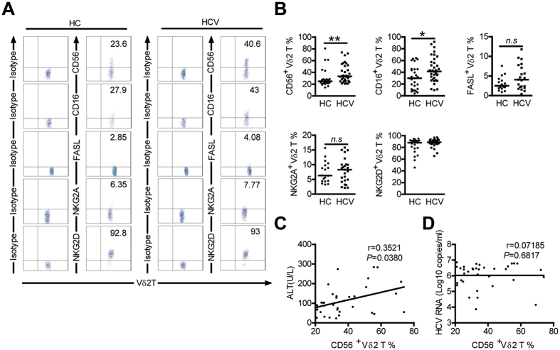
Figure 2. Comparison of the expression of NK cell-associated antigens on Vδ2 T cells in HCs and HCV-infected patients.
(A) Representative dot plots show the expression of CD56, CD16, FasL,NKG2A and NKG2D on Vδ2 T cells from HCs and HCV-infected patients. (B) Frequency of Vδ2 T cells displaying the indicated surface markers in HCs and HCV-infected patients. CD56, n = 25 and 35, respectively; CD16, n = 27 and 36, respectively; FasL, n = 19 and 21, respectively; NKG2A n = 18 and 25, respectively; NKG2D, n = 21 and 32, respectively. Horizontal lines indicate the median. (C,D) Correlation analysis of the percentages of CD56 + Vδ2 T cells and the serum ALT levels (C) or HCV RNA loads (D) in HCV-infected patients. n = 35. *p < 0.05, **p < 0.01.

