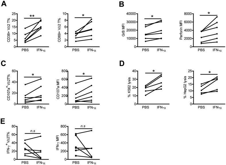
Figure 6. Vδ2 T cells of HCs are activated and upregulate cytolytic activity but not IFN-γ production after in vitro exposure to IFN-α.
(A–C,E) PBMCs from HCs were preincubated with or without IFN-α for 24 h. (A,B) Expression of activation markers CD38 and CD69 (A), and cytolytic enzymes GrB and perforin (B) on Vδ2 T cells was assessed by flow cytometry. (C,E) Expression of CD107a (C) and IFN-γ (E) on Vδ2 T cells upon zoledronate stimulation was analyzed by flow cytometry. n = 6 for each group. (D) Percentages of K562 and HepG2 lysis byVδ2T cell lines preincubated with or without IFN-α for 24 h. n = 5 for each group. *p < 0.05, **p < 0.01.

