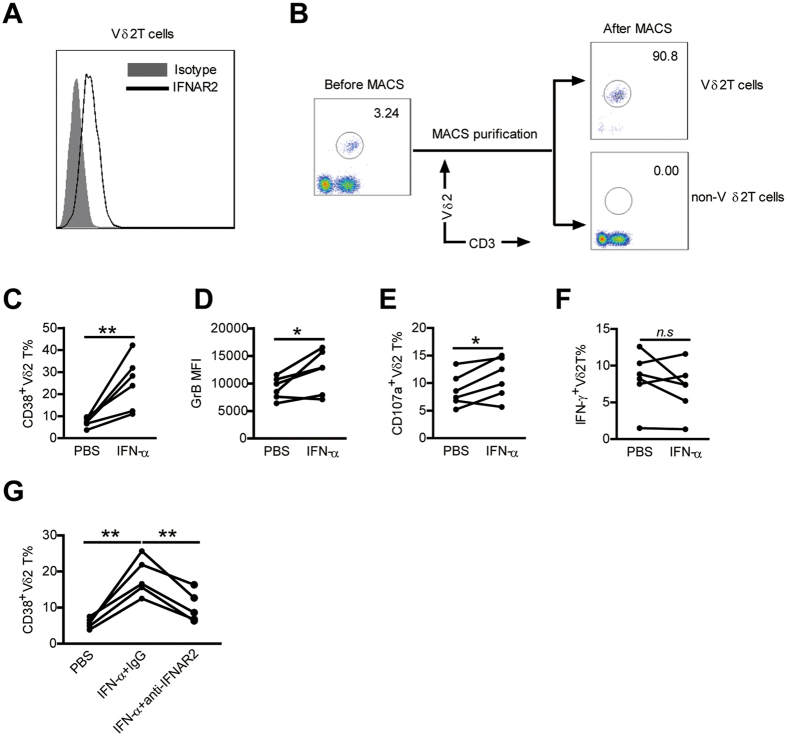
Figure 7. IFN-α could directly modulate Vδ2 T-cell phenotype and function.
(A) PBMCs from HCs were stained with an unlabeled primary antibody against IFNAR2 or an isotype-matched control antibody (mouse IgG2a), and then by a phycoerythrin (PE)-conjugated anti- mouse IgG2a secondary antibody. IFNAR2 expression on Vδ2 T cells was analyzed. A representative staining profile is shown. (B) PBMCs of healthy blood donors were separated for Vδ2 T cells and non- Vδ2 T cells using magnetic beads. Purified cells were stained with anti- Vδ2 T mAb and anti-CD3 mAb and analyzed by flow cytometry. The representative dot plots from six experiments are shown. (C–F) Purified Vδ2 T cells were preincubated with or without IFN-α for 24 h. (C,D) Expression of activation markers CD38 (C), and cytolytic enzymes GrB (D) on Vδ2 T cells was assessed by flow cytometry. (E,F) Expression of CD107a (E) and IFN-γ (F) on Vδ2 T cells upon zoledronate stimulation was analyzed by flow cytometry. n = 6 for each group. (G) Purified Vδ2 T cells were cultured in medium containing PBS or 100 U ⁄ml IFN-α, a blocking antibody against type I IFN receptor (anti-IFNAR2, 5 μg ⁄ml) or an isotype control antibody was added to the medium. After 24 hr of culture, the expression of CD38 on Vδ2 T cells was assessed. n = 5. *p < 0.05, **p < 0.01.

