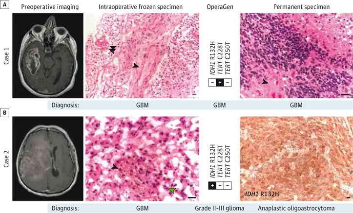
Figure 3. Sensitive Detection of Glioma-Specific Somatic Variants Within an Intraoperative Timeframe Using Operative Genotyping (OperaGen).

A, Case 1 demonstrates the detection of TERT promoter mutation from a specimen that was diagnosed as high-grade glioma on intraoperative frozen specimen analysis (single arrowhead indicating microvascular proliferation; double arrowhead, necrosis) and GBM on permanent specimen analysis (single arrowhead indicating microvascular proliferation). Both specimens stained with hematoxylin-eosin. B, Case 2 frozen specimen (hematoxylin-eosin) was diagnosed as GBM based on the presence of microvascular proliferation (single black arrowhead) and mitotic figures (single green arrowhead). The OperaGen detection of IDH1 R132H would alternatively have been consistent with WHO grade II or III glioma, which was in agreement with findings of the permanent specimen analysis of IDH1-mutant anaplastic oligoastrocytoma (immunohistochemically stained). All scale bars represent 10 μm. GBM indicates glioblastoma; WHO, World Health Organization.
