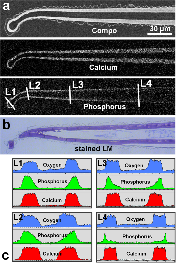
Figure 3. Analyses of a median longitudinal section through an embedded Loasa pallida stinging hair.
(a) The SEM image of the sectioned block face shows the high mineral content in the wall by the compositional contrast of the BSE image. The element mapping images show the Ca and P distribution. (b) The light microscopy image of a toluidine blue-stained thin section (not exactly in the median plane) indicates that the entire cell wall contains organic material in addition to the mineral components. (c) EDX line scans show the concentration profiles for Ca, P, and O at different positions (L1 to L4). Note the continuous decrease of the phosphorus concentration from the outside towards the inner wall (line scan L3).

