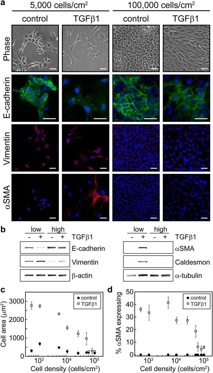
Figure 1. Increasing cell density blocks TGFβ1-induced EMT.
(a) Phase contrast microscopy images of NMuMG cells and immunofluorescence staining of EMT markers at seeding densities of 5,000 cells/cm2 and 100,000 cells/cm2 with and without TGFβ1 treatment. Blue stain shows cell nuclei. Scale bars: 50 μm. (b) Western blot analysis of EMT markers for cells seeded at low (5,000 cells/cm2) and high (100,000 cells/cm2) densities with and without TGFβ1. (c) Mean cell area as a function of cell seeding density. (d) Percentage of cells expressing αSMA as a function of cell seeding density.

