Figure 1.
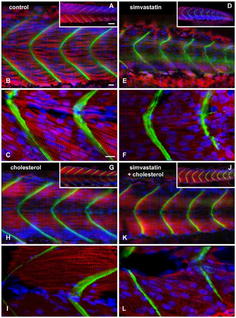
Structural analysis of mild phenotype, and its rescue with cholesterol. Confocal analysis of zebrafish embryos at 48 hours post-fertilization stained with sarcomeric alpha-actinin (red), laminin (green) and DAPI (blue). In the control embryo, a single slice shows eight regularly sized somites with straight angle septa (A). In the 0.3 nM simvastatin-treated embryo (D), there is a weak alpha-actinin stain, showing altered somites. In embryos treated with cholesterol alone (G), the somites appear regular in size. In embryos treated with simvastatin and cholesterol (J), the sizes of the somites are more similar to that of the control embryos than the embryos treated with simvastatin alone, although the septa are still irregular. In a projected region of six sequential slices (B, E, H and K), we can appreciate the continuity of the septa in the control embryos (B) and the embryos treated with cholesterol alone (H), whereas, in the simvastatin group, the septa is discontinuous and irregular (E). Simvastatin embryos rescued with cholesterol (K) show a more continuous septa than embryos treated with simvastatin alone, although these patterns are still not as regular as that in the control. At higher magnification (C, F, I and L), the striations are clearly seen in the straight myofibrils in control embryos (C) and embryos treated with cholesterol alone (H). In simvastatin-treated embryos (F), the myofibrils are shorter, curved and have a reduced stain for alpha-actinin. Embryos treated with simvastatin and cholesterol (L) have more alpha-actinin in striated myofibrils than simvastatin alone. Scale (A, D, G, J), 50 μm; (B, C, E, F, H, I, K, L), 10 μm.

