Figure 3.
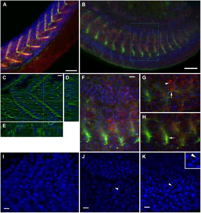
Structural analysis of severe phenotype: Intermediate filaments and adhesion complexes. Confocal analysis of zebrafish embryos at 24 hours post-fertilization (hpf) stained with desmin (green), vinculin (red) and DAPI (blue). Control embryo shows septa in chevron and large, regularly spaced somites in the stack projection (A). In the confocal projection of a 0.75 μM simvastatin-treated embryo (b), somites are shorter and septa labeling with vinculin is fainter, as is the myofibrillar desmin stain. In some regions, there is a small accumulation of desmin around the remnants of a septa (B). Analyzed in more detail, a selected slice of a control embryo shows the distribution of desmin around the nuclei, which are regularly spaced, and around the septa (C). The orthogonal sections show the septa (D) and a myofibril (E). In a detail of the inset in (B), the confocal projection highlights the concentration of desmin and vinculin staining (f). A selected slice (g) of the previous stack show a thin myofibril stained for desmin (arrow) close to an aggregate of vinculin (arrowhead). Another selected slice (h) shows the desmin accumulation in the remainder of the septum (arrowhead). In a projection of a DAPI labeling of control embryos (I), the gap where the septa lie is clearly visible. In the projections of treated embryos (J, K), the gap between the nuclei is not visible, and some apoptotic nuclei can be seen (arrowheads). This is clearer in the magnified inset in (K). Scale (A, B) 50 μm; (C, F, I, J, K) 10 μm.

