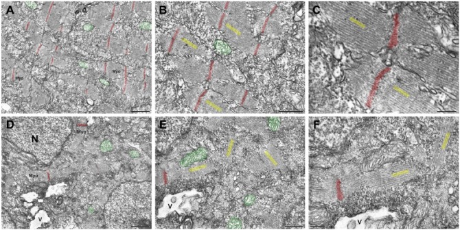
Figure 5.
Transmission electron microscopy of zebrafish embryos treated with simvastatin. The untreated embryos (A–C) present organized bundles of myofibrils (Myo), and exhibit well-preserved mitochondria (green). Note that the Z lines (red) are aligned to each other and periodically spaced. The myofibrils around a Z-line are better visualized in figures (B, C), and their direction is indicated in yellow, showing their alignment. In the simvastatin-treated embryos (D–F), myofibrils (Myo) are disorganized and the alignment of Z lines is lost. Vacuoles (V) are observed, as well as several mitochondria (green) and nuclei (N). At a higher magnification (E, F), the myofibrils are clearly oriented in several directions (yellow). Note that, even when though the myofibrils are not aligned, the thick and thin filaments, marked in yellow within each myofibril, are parallel to each other, as happens in control. Scale (A, D) 1 μm; (B, E) 500 nm; (C), 200 nm; (F), 250 nm.

