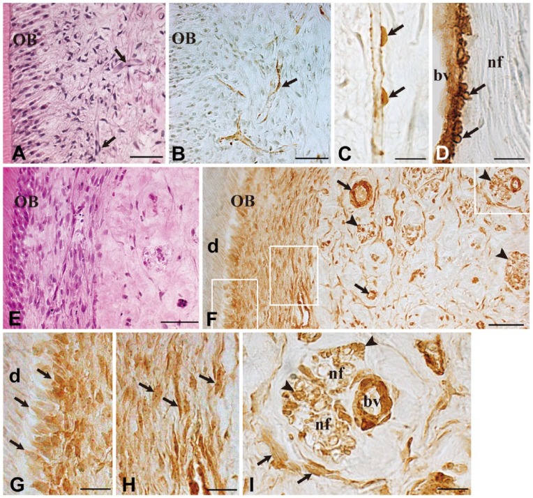
Figure 2.
Immunohistochemical staining for α-SMA in uncultured normal pulp and after 7 days of culture. Hematoxylin-eosin staining (A, E) and immunostaining for α-SMA (B–D, F–I) in uncultured normal tissue (A–D) and after 7 days of culture (E–I). Higher magnification views of the boxed areas in (F) are shown in (G), (H) and (I), respectively. In uncultured normal pulp, capillaries are observed in the periphery of the dental pulp (A, arrows), and immunoreactions for α-SMA are obvious in pericytes (B and C, arrows) and vascular smooth muscle cells (D, arrows). After 7 days of culture, α-SMA immunoreactivity can be recognized in various cell types (F, arrows: blood vessels; arrowheads: nerve fibers) (F) including odontoblasts (G, arrows), fibroblasts (H and I, arrows), and Schwann cells (I, arrowheads). OB, odontoblast; d, dentin; bv, blood vessel; nf, nerve fiber. Scale (A–B, E–F) 50 µm; (C–D, G–I) 20 µm.

