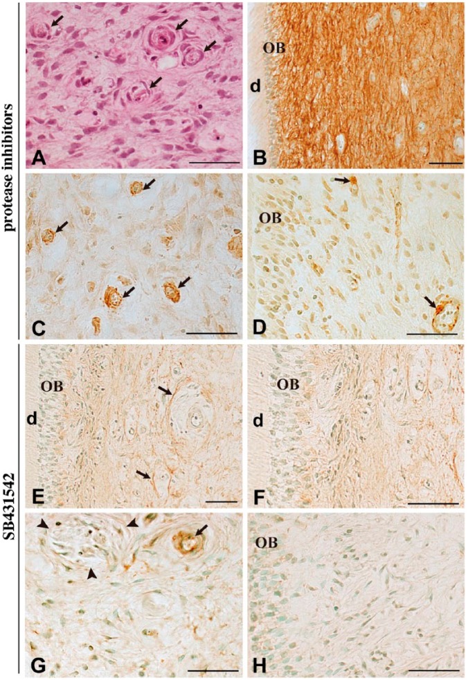
Figure 3.
Immunohistochemical staining for fibrillin-1 and α-SMA after 7 days of culture in the presence of protease inhibitors (4-Abz-Gly-Pro-D-Leu-D-Ala-NH-OH and P1860) or TGF-β type I receptor inhibitor (SB431542). Hematoxylin-eosin staining (A) and immunostaining for fibrillin-1 (B, E–F) and α-SMA (C, D). The tissue morphology in the presence of protease inhibitors appears to be maintained, even in blood vessels (A, arrows). Administration of protease inhibitors prevents the loss of fibrillin-1 immunoreactivity (B) and little immunoreactivity for α-SMA is noted in fibroblasts (C) and odontoblasts (D). Cells along the blood vessels remain positive for α-SMA (C–D, arrows). In the presence of SB431542, fibrillin-1 staining becomes rather faint except around the blood vessels (E, arrows), and the interodontoblastic area is devoid of fibrillin-1 (F). Most dental pulp cells are negative for α-SMA (G–H, arrowheads in G: nerve fiber), except along the blood vessel (G, arrow). OB, odontoblast; d, dentin. Scale, 50 µm.

