Figure 2.

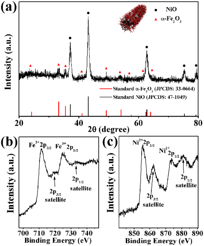
(a) XRD pattern of α-Fe2O3/NiO nanotubes. (b,c) XPS patterns of the Fe 2p and Ni 2p regions of the α-Fe2O3/NiO nanotubes, respectively.

(a) XRD pattern of α-Fe2O3/NiO nanotubes. (b,c) XPS patterns of the Fe 2p and Ni 2p regions of the α-Fe2O3/NiO nanotubes, respectively.