Figure 2.
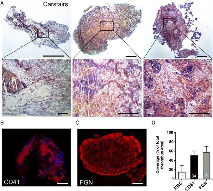
Histological analysis of thrombus aspirated from intracoronary stents. (A) Representative Carstairs stainings of thrombus aspirates (n = 11). Upper row: overview image (left bar, 50 µm; other bars 100 µm). Bottom row: insets of the overview images (left bar, 25 µm; other bars 50 µm); platelets are stained in grey blue to navy, fibrin/fibrinogen in red and erythrocytes (RBC) in yellow; (B) representative image of platelet aggregation area (CD41 positive) in thrombus aspirates (n = 7). Nuclei were counterstained with Hoechst. Bar, 100 µm; (C) fibrin/fibrinogen immunofluorescence staining (n = 34). Nuclei were counterstained with Hoechst. Bar, 100 µm; (D) Coverage of whole thrombus area (%) with RBC, platelet (CD41) and fibrin/fibrinogen rich areas. Due to co-localization, overall coverage exceeds 100%; data are shown as mean + SD.

