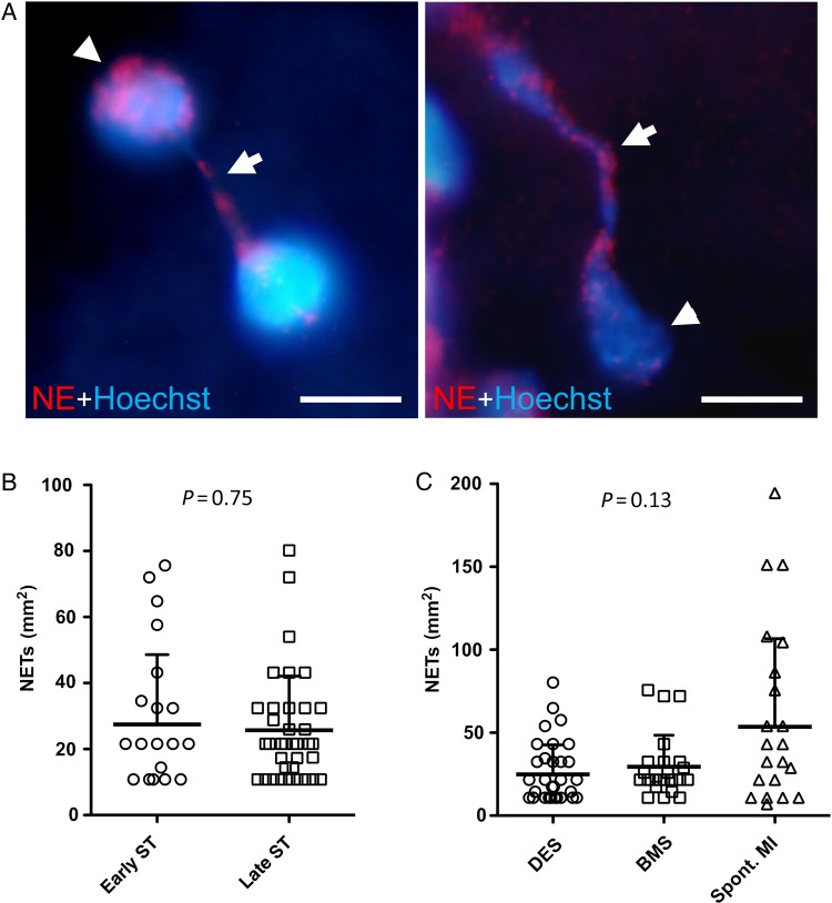
Figure 4.
Detection of neutrophil extracellular traps in stent thrombus specimens. (A) Immunofluorescence images of neutrophil extracellular traps stained for neutrophil elastase and DNA (Hoechst). Extracellular DNA originates from neutrophil elastase positive neutrophils. Arrowheads, nuclei; arrows, neutrophil extracellular trap fibres. Bars, 5 µm; (B) number of neutrophil extracellular traps in early (n = 23) vs. late (n = 37) stent thrombosis (P = 0.75); (C) quantification of neutrophil extracellular traps in thrombi derived from drug-eluting stents (n = 36), bare metal stents (n = 23), and spontaneous myocardial infarction (spont. myocardial infarction) (n = 25) (P = 0.13); data are shown as mean + SD, each symbol in (B) and (C) represents one individual patient.

