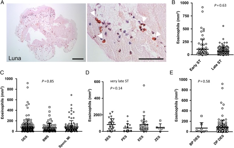
Figure 5.
Eosinophil accumulation in stent thrombus specimens. (A) Eosinophils in human thrombi were identified by Luna staining. Arrowheads indicate eosinophils (red-brown colour). Bars, 100 µm (left) and 50 µm (right); (B) Number of eosinophils in early (n = 71) vs. late (n = 146) stent thrombosis (P = 0.63); (C) Quantification of eosinophils in thrombi derived from drug-eluting stents (n = 143), bare metal stents (n = 66) and spont. myocardial infarction (n = 93) (P = 0.85); (D) Number of eosinophils in very late ST according to drug-eluting stents type; SES (n = 24), PES (n = 16), EES (n = 27), ZES (n = 6) (P = 0.14); (E) Number of eosinophils according to drug-eluting stents polymer type; bioabsorbable polymer (BP drug-eluting stents): n = 8, durable polymer (DP drug-eluting stents): n = 120 (P = 0.58); data are shown as mean + SD, each symbol in (B–E) represents one individual patient.

