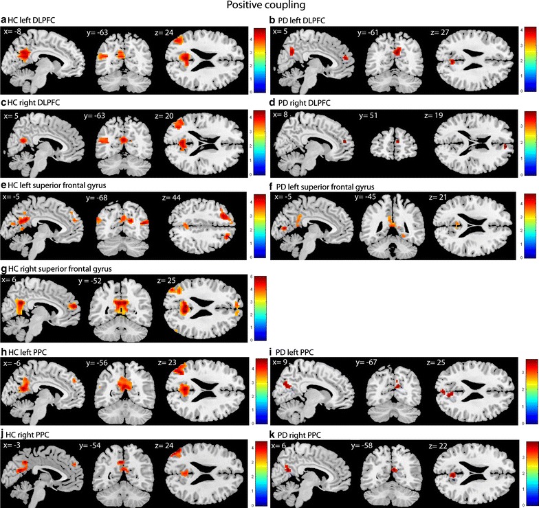
Fig. 1.

Positive coupling of the DLPFC, superior frontal gyrus and PPC in HC and PD. T-statistic images of positive connectivity in the [successful shift > successful repeat] contrast, corrected for mean RT on shift trials. A voxel-level threshold of p < .001 is used with an extent threshold of 10 voxels. The images are overlaid on ch2better MNI template with MRIcron, coordinates are in MNI space. The coloured bar indicates the Z-value
