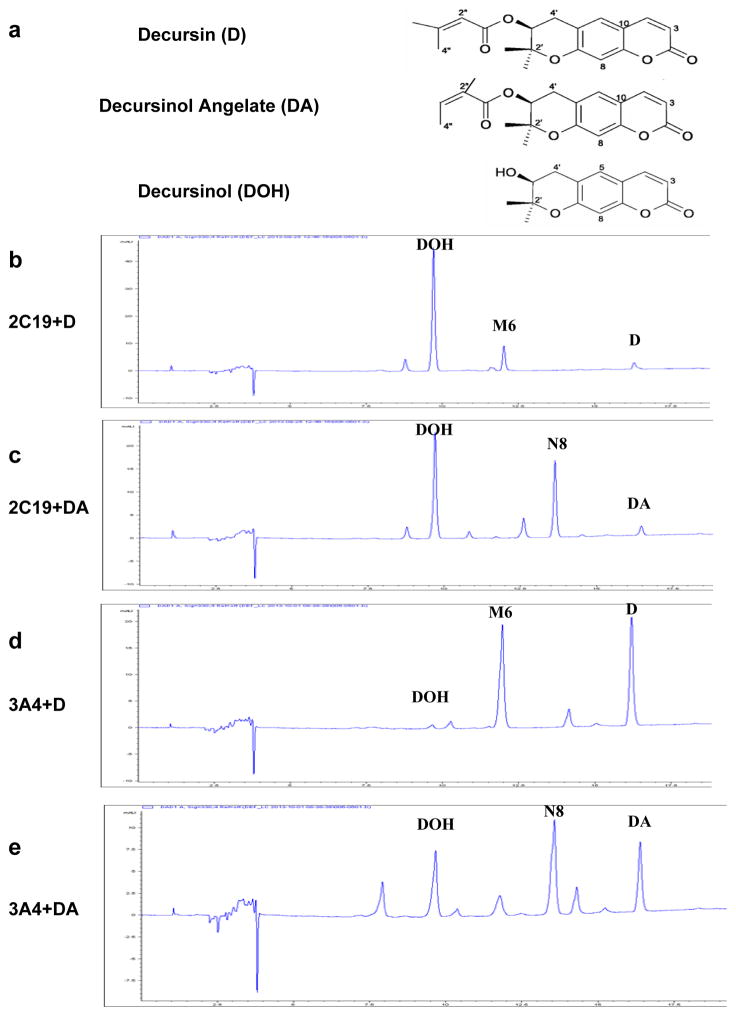
Figure 1.
In vitro metabolism of D and DA by recombinant human CYP 2C19 and 3A4 proteins. Panel a, Structures of decursin (D), decursinol angelate (DA) and decursinol (DOH). Panel b, CYP 2C19 metabolism of D. Panel c, CYP 2C19 metabolism of DA. Each reaction system contained 10 pmol/mL 2C19. Panel d, CYP 3A4 metabolism of D. Panel e, CYP 3A4 metabolism of DA. Reaction system contained 100 pmol/mL 3A4. Reactions were terminated after 10 minutes. UV detection wavelength was 330nm for HPLC-chromatograms. M6 and N8 correspond to respective mono-oxygenated metabolite of D and DA in human liver microsome incubation we previously identified (Li et al., 2013a).

