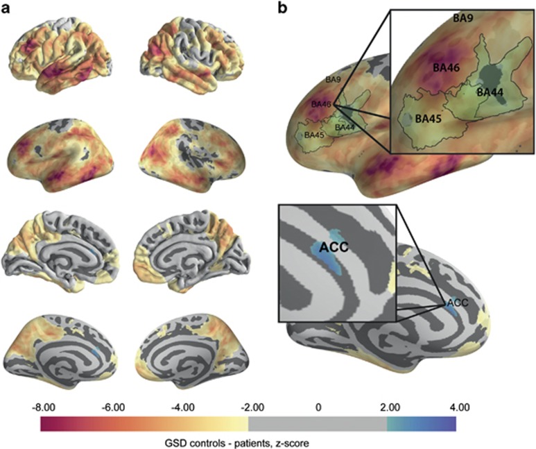
Figure 3.
Per-vertex comparison of gyral–sulcal thickness differences (GSDs). (a) Local GSDs are regionally increased particularly in dorsolateral prefrontal cortex (DLPFC/BA46), superior temporal gyrus, inferior temporal gyrus and inferior parietal gyrus. (b) The regional pattern of sulcal-specific thinning is consistent with neuropathology studies of schizophrenia, which have identified layer II thinning in BA46,13 but not BA9, BA44 (ref. 39) or anterior cingulate cortex.38

