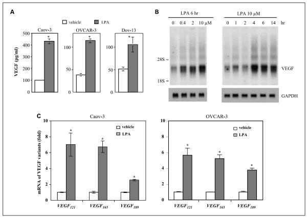
Fig. 1.
Induction of multiple isoforms of VEGF by LPA in ovarian cancer cell lines. A, LPA-stimulated secretion of VEGF. Caov-3, OVCAR-3, and Dov-13 cells were plated in 6-well plates, cultured to ~60% confluence, starved for 24 h and then stimulated with LPA (10 μmol/L) or vehicle (BSA control) for 16 h. VEGF levels (pg/mL) in culture supernatants were quantified using VEGF ELISA kits. B, LPA-induced VEGF mRNA expression. Caov-3 cells were starved and treated with LPA for 6 h at indicated concentrations (left) or treated with 10 μmol/L LPA for different periods (h; right). Total cellular RNA was isolated and analyzed by Northern blotting using 32P-labeled VEGF cDNA as a probe. The locations of 28S and 18S RNA were indicated. The membrane was reprobed with GAPDH to confirm comparable loading among samples. C, Caov-3 and OVCAR-3 cells were treated with 10 μmol/L LPA for 4 h before RNA preparation. The mRNA levels of three major VEGF variants (VEGF121, VEGF165, and VEGF189) were determined by quantitative PCR using quantitative PCR primers specific for each of the VEGF isoform as detailed in Materials and Methods. For this and the following illustrations, all numerical data are mean ± SD, representative of three independent assays. *, P < 0.05, statistical significant differences.

