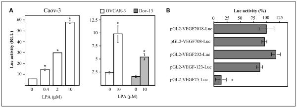
Fig. 3.
Activation of VEGF transcription by LPA. A, LPA-stimulated activation of the VEGF promoter. Caov-3, OVCAR-3, or Dov-13 cells were transfected with pGL2-VEGF2018-Luc, starved for 1 day, and stimulated with LPA for 6 h at the indicated concentrations. The luciferase activities were analyzed with luciferase assay kits (Promega). Results were presented as relative light units (RLU). B, VEGF promoter sequences responsible for LPA stimulation were mapped to a 123-bp fragment proximal to the initiation site. Caov-3 cells were transfected with a series of deletion mutants containing 2018, 708, 232, 123, or 25 bp fragments of the VEGF promoter. LPA-stimulated luciferase activity was determined as in A. Data are presented as relative percentages with the activity of cells transfected with pGL2-VEGF2018-Luc defined as 100%. In this and the following figures, luciferase activities were normalized to β-galactosidase activity in the cells cotransfected with pCMVβ-gal.

