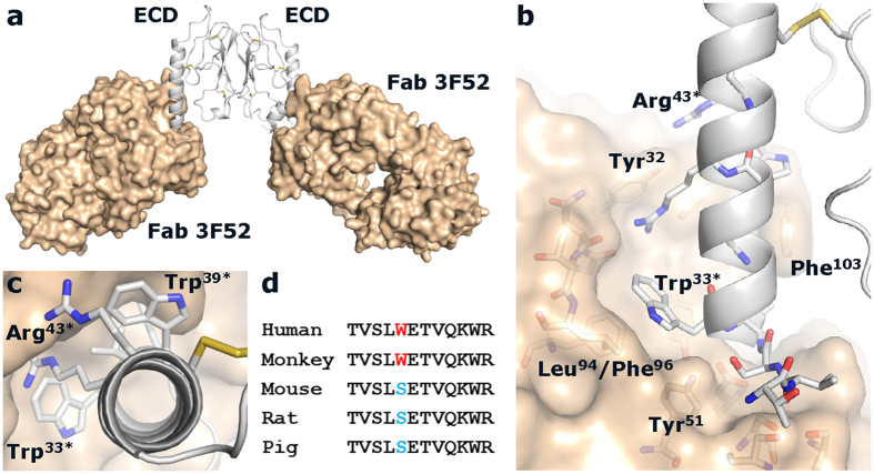
Figure 3. Crystal structure of the GLP-1R ECD/Fab 3F52 complex.
The Fab 3F52/ECD complex was crystallized with two complexes in the asymmetric unit as shown in (a). The GLP-1R ECD is shown in grey as a ribbon illustration and Fab 3F52 is shown in gold as a surface illustration. (b) Zoom in on the Fab-receptor interface showing selected residues at the receptor/Fab interface in sticks. (c) Top-down view of the α-helix of the GLP-1R ECD. (d) Sequence alignment of the Fab 3F52 epitope of GLP-1R from different species.

