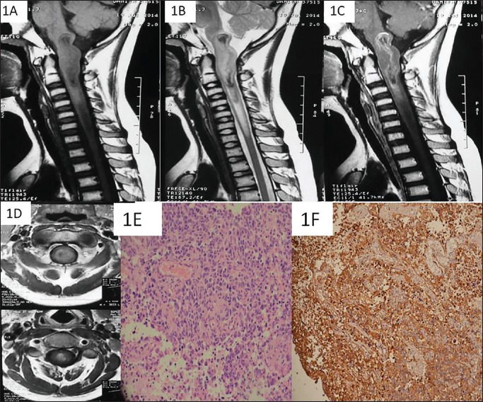
Figure 1.
On MRI of the cervicomedullary junction, a T1 (a) and T2 (b) heterogenous mass is seen expanding the lower medulla and the upper cervical spinal cord till the level of C3. The mass has hypo intense areas on both T1/T2 images (a and b). On contrast image, there is peripheral rim like enhancement of the mass without any enhancement of the centre (c). The mass is almost centrally located inside the neuraxis (d). The tumor showed pleomorphic cells displaying hyper chromatic nuclei, scant cytoplasm with brisk mitosis on H & E staining using 40x magnification (e). There was a strong membraneous positivity noted for CD99 (f)

