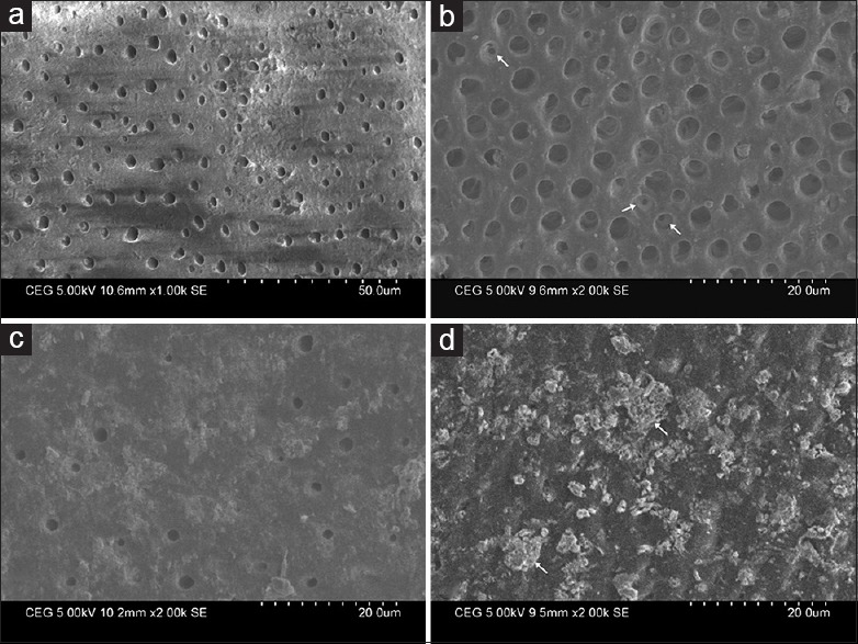
Figure 1.

Scanning electron micrographs of specimens treated with (a) 17% ethylenediaminetetraacetic acid showing open dentinal tubules, (b) 2% sodium fluoride showing partial occlusion of dentinal tubules (arrows), (c) nano-hydroxyapatite showing a predominantly higher number of tubular occlusion and partial coverage of the dentinal surface with film or precipitate, (d) combination of nano-hydroxyapatite and 2% sodium fluoride showing complete occlusion of all the dentinal tubules; presence of a protective film and agglomerated precipitates (arrows) on the surface
