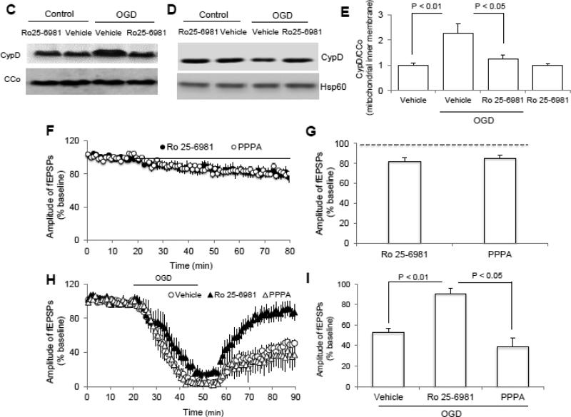
Figure 4. N-methyl D-aspartate receptor subunit 2B (NR2B) activation is involved in OGD-induced cyclophilin D (CypD) translocation and synaptic injury.
A-B) Representative immunoblotting bands (A) and Quantification of (B) show the phosphorylation level of NR2B at ser1303 in indicated groups. C-D) Representative immunoblotting bands show CypD levels in mitochondrial inner membrane fraction and matrix (D) in indicated groups. E) Quantification of CypD immunoreactive bands normalized to CCo in indicated groups shown in panel C. N =4 mice per group. F) Inhibition of either NR2A (PPPA, 0.5 μM) or NR2B (Ro 25-6981, 1 μM) by its inhibitor perfusion (bar) suppressed synaptic transmission under normal condition. G) Average of the last 5 min of reperfusion fEPSPs amplitude in the indicated groups. H) Inhibition of NR2B but not NR2A significantly ameliorated synaptic injury after OGD. I) Synaptic transmission recovery of field-excitatory post-synaptic potentials (fEPSPs) calculated as the averaged relative amplitude of fEPSPs compared to baseline values after re-introduction of oxygenated normal artificial cerebrospinal fluid (ACSF, see methods section) (from 35 to 40 min after the end of OGD). N = 6-10 slices from 4-5 male mice (3-4 month-old age) per group.


