
Figure 8.
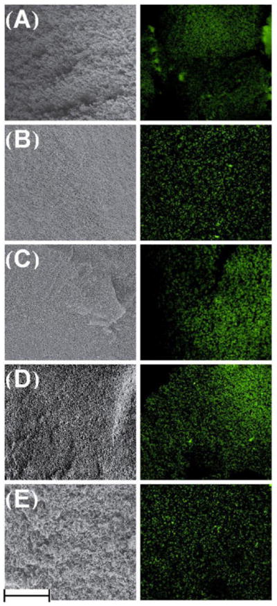
SEM (left) and confocal microscopy (right) images, showing the gels in dry and wet environments, respectively. These correlated images demonstrate that the gel microstructure retained after hydration. Confocal imaging was performed by illuminating pores with Rhodamine 123. (A) Control; (B) Pre48CX; (C) Pre24CX; (D) CCX; (E) Post24CX. Scale bar equals 100 μm.