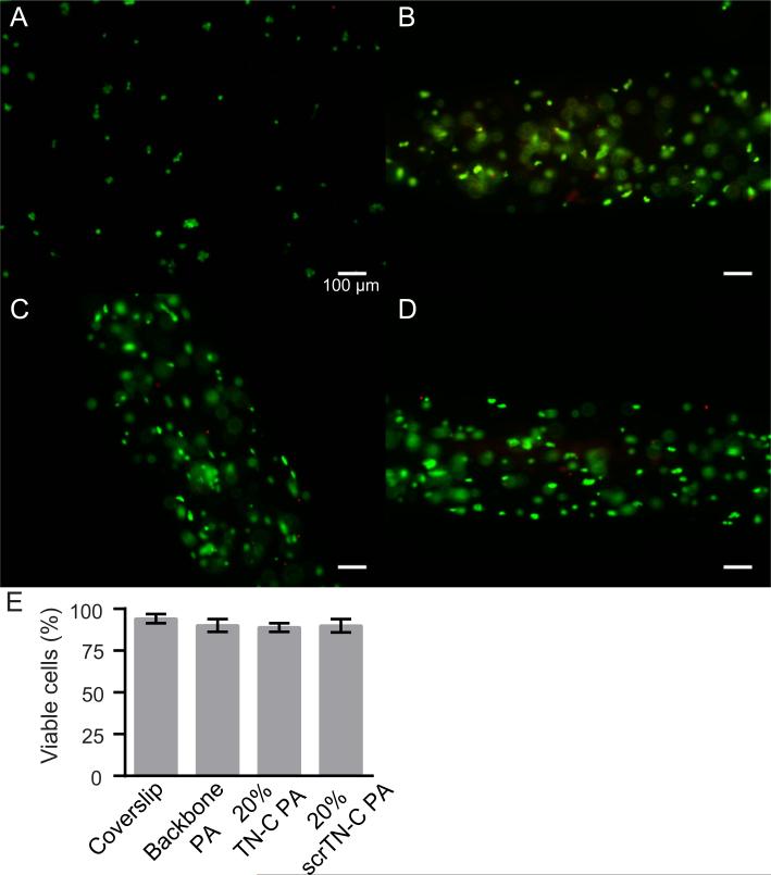
Fig. 3.
Cell viability in PA gels after two days of culture. (A) P19-derived neurons on a PDL/laminin coverslip, (B) in backbone PA gel, (C) in backbone PA and 20% TN-C PA gel, and (D) in backbone PA and 20% scrTN-C PA gel. Living cells are labeled green (calcein-AM) and dead cells are labeled red (propidium iodide). Scale bars: 100 μm. (E) The percentage of cells that were viable for each condition.

