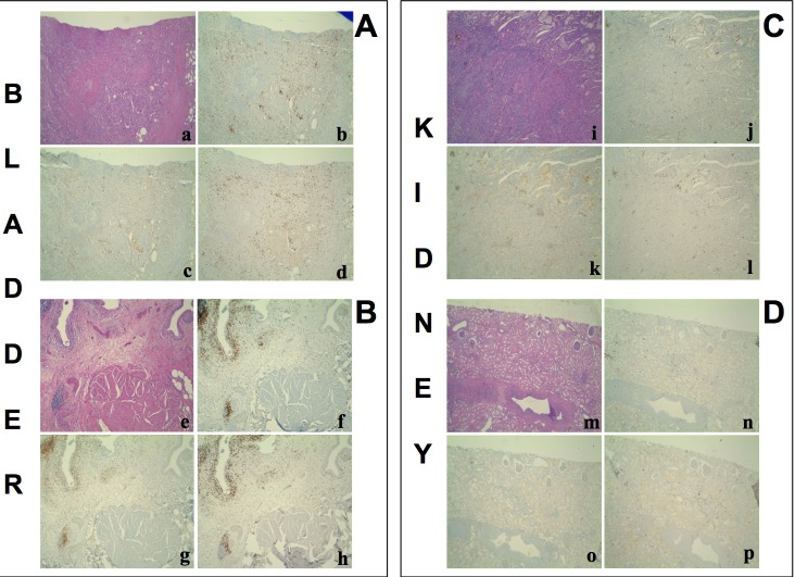
Figure 3. Hematoxylin-Eosin stain and immunohistochemistry for CD3+, CD4+, CD8+ T cells on.
A. a poorly differentiated urothelial carcinoma of the bladder B., the autologous histologically free-of-tumor bladder tissue (both from patient #1), C. a papillary type II renal cell carcinoma and D. the autologous histologically free-of-tumor cortical renal tissue (both from patient #30). a, e, i, m: Haematoxylin-Eosin stain (4x magnification); b, f, j, n: immunohistochemistry using an anti-CD3 antibody (4x magnification); c, g, k, o: immunohistochemistry using an anti-CD4 antibody (4x magnification); d, h, l, p: immunohistochemistry using an anti-CD8 antibody (4x magnification). a-h: an intense T lymphocyte infiltrate is shown both in the tumour and the histologically free-of-tumor mucosa; lymphocytes are sparse or in nodular aggregates and localize between neoplastic cells as well as in the interstitium. In non-neoplastic mucosa a comparable infiltrate is seen both in the intraepithelial tissue and in the subepithelial connective tissue. i-p: While the tumor shows an evident T lymphocyte infiltrate, rare T lymphocytes are present in the non-neoplastic epithelium.

