
Figure 6. Orthotopic implants of CXCR2 knock-down cells demonstrate inhibited proliferation of tumor cells and increased fibrosis.
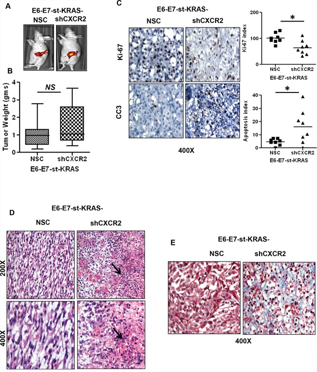
A. In vivo GFP images of E6-E7-st-KRAS-NSC or E6-E7-st-KRAS-shCXCR2 cells implanted orthotopically in the pancreas of nude mice. B. The mean weight of tumors derived from E6-E7-st-KRAS-NSC or E6-E7-st-KRAS-shCXCR2 cells. C. Representative images of immunohistochemistry (IHC) and quantified stain score for Ki-67 and cleaved caspase 3 (CC3). IHCs were quantified as the average of positive cells in five independent fields per tumor at 400X. D. H&E staining demonstrating infiltration of leukocytes in tumors derived from mice bearing either E6-E7-st-KRAS-NSC or E6-E7-st-KRAS-shCXCR2 cells. E. Masson's trichrome stain showing collagen deposition. Statistical significance determined by paired Student's t-test (*p ≤ 0.05, **p ≤ 0.01, ***p ≤ 0.001, NS p > 0.05).