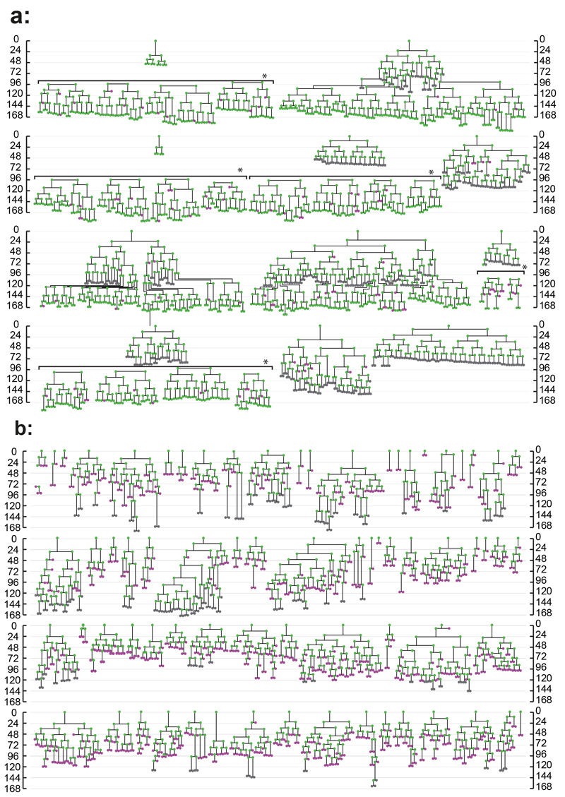
Figure 2. Lineage trees of Neonatal Foreskin Keratinocytes cultured at clonal density.
Scale indicates time since plating in hours. Magenta indicates cells that did not divide within 48 hours, green cells which were observed to divide and grey cells those which could not be tracked for at least 48 hours. Horizontal brackets in a, marked by *, indicate representative cells tracked within a single colony. a: expanding trees, b: balanced trees, see text for details. A total of 81 trees from 3 independent experiments is shown.

