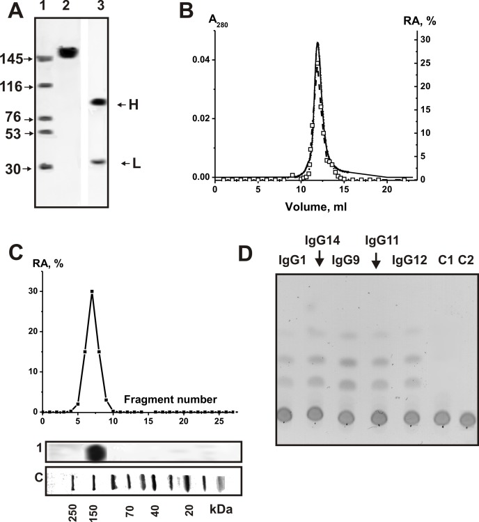
Fig 1.
SDS-PAGE analysis of homogeneity of csf-IgGmix (7 μg; lanes 2 and 3) corresponding to 15 CSFs of MS patients in 3–16% gradient gel before (lane 2) and after treatment with DTT (lane 4) followed by silver staining (A). Lanes 2 and 3 correspond to Western-blotting; Abs against human amylase were used in the case of IgG (lane 2) and human amylase (lane 3). The arrows (lane SP) indicate the positions of molecular mass markers. FPLC gel filtration of csf-IgGmix on a Superdex 200 column in an acidic buffer (pH 2.6) destroying immunocomplexes after Abs incubation in the same buffer (B): (—), absorbance at 280 nm (A280); (□), relative activity (RA) of IgGs in the hydrolysis of MHO. A complete hydrolysis of MHO was taken for 100%. In-gel assay of MHO-hydrolyzing activity of csf-IgGmix (■; 15 μg) of MS patients. The relative MHO -hydrolyzing activity (RA, %) was revealed using the extracts of 2-3-mm fragments of one longitudinal slice of the gel. The RA of IgGs corresponding to complete hydrolysis of MHO was taken for 100%. The second control longitudinal slice of the same gel was stained with Coomassie Blue (lane 1). Lane C shows positions of protein markers. TLC analysis of the hydrolysis of MHO by IgGs from CSFs of different MS patients (D). MHO (1.67 mM) was incubated for 6 h at 37°C without Abs (lanes C2) and in the presence of 0.07 mg/ml IgGmix from the plasma of healthy donors (lane C1) as well as individual IgGs (0.025 mg/ml, 2 h) from CSFs of different MS patients (lanes 1–5). The average error in the initial rate determination from three experiments did not exceed 7–10%. For details, see Materials and methods.

