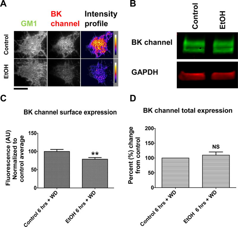
Figure 9.

The BK channel remains internalized after 6-hrs of EtOH exposure followed by 18-hrs of withdrawal. Hippocampal neurons were incubated with EtOH for 6-hrs, washed and left in withdrawal 18-hrs. Neurons were fixed (or lysed for Western blot) and stained immediately against the BK channel afterwards. (A) Representative images showing a reduction in BK perimembrane expression after 6-hrs of EtOH exposure plus 18-hrs of withdrawal when compared to control. Scale bar = 10μm (B) Representative blots showing no change in total levels of BK protein expression in neurons after 6-hrs of EtOH exposure plus 18-hrs of withdrawal when compared to control. (C) Perimembrane expression of the BK channel quantified after TIRF imaging is significantly reduced after EtOH exposure followed by withdrawal when compared to control, unpaired t-test, p < 0.01. Error bar is ±SEM, (control N=26, EtOH N=29 neurons) (D) Total BK channel protein levels quantified via Western blot show not significant difference when compared to control group, unpaired t-test, p > 0.05 (N=4).
