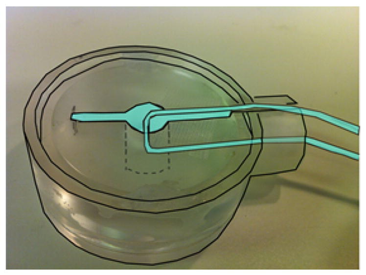
Fig. 3.

Ionic current phantom used to estimate temporal standard deviation in the phase data. Cyan-colored areas are filled with 0.9% saline containing 5 mM CuSO4. The 100 μA current was restricted to the glass capillary tube. Dimensions of chamber are same as in the cerebellum study.
