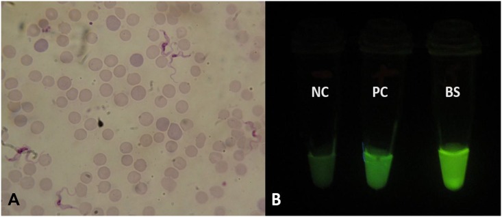
Fig 1. Microscopic and LAMP fluorescent visualisation of trypanosomes.
Detection of trypanosomes in patient’s blood by (A) microscopy (×100 magnification) and (B) loop-mediated isothermal amplification (LAMP). Visual appearance of results for human serum resistance-associated gene (SRA)-LAMP of a patient with Early Stage human African trypanosomiasis (HAT). Loopamp Fluorescent detection reagent was added to the reaction mixture at the beginning of the assay. The reactions were incubated at 64°C for 30 minutes. In contrast to the light green background fluorescence in the negative samples, positive samples exhibit a bright fluorescent green colour when visualized under the transilluminator. NC: Negative control (distilled water); PC: Positive control (Trypanosoma brucei rhodesiense); BS: patient blood sample.

