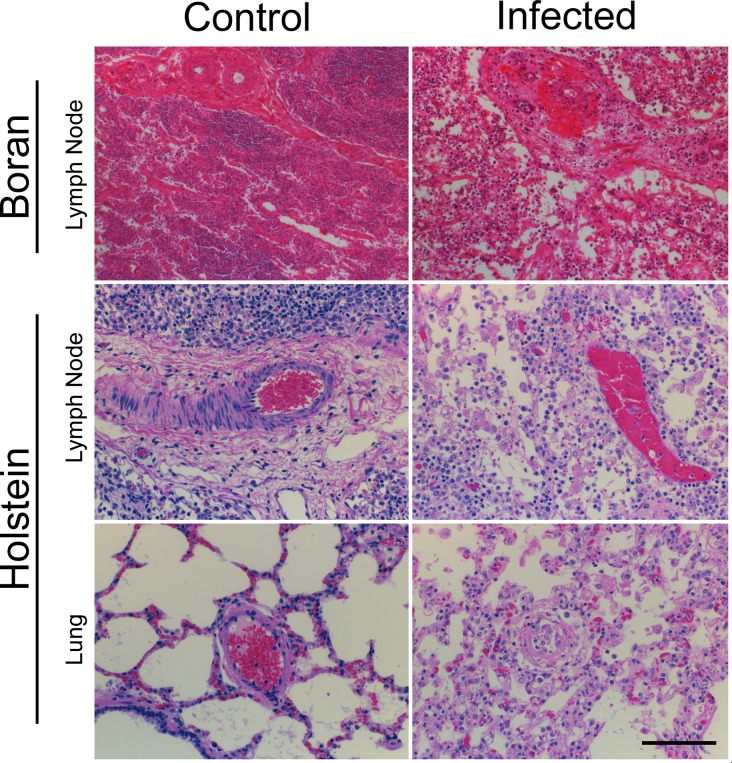
Fig 1. Histologic Lesions in Holstein and Boran cattle.
Representative photomicrographs of H&E stained sections of lymph node medulla in uninfected control and infected Boran (day 12) and Holstein calves, and lung from uninfected control and infected Holstein calves. Note: Severe disruption of vessel walls by mononuclear cells and fibrinoid degeneration (lung and lymph node), and lymphohistiocytic interstitial pneumonia with edema (lung). Scale bar: 200 μm.

