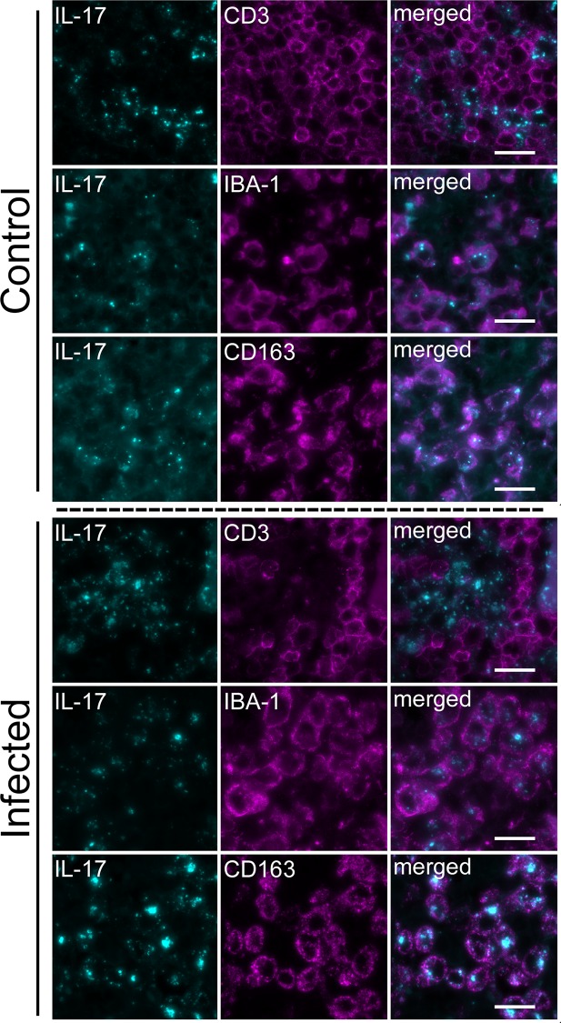
Fig 4. Dual Fluorescence Labeling of Leukocytes in Lymph Nodes.
Shown are the results of dual fluorescence labeling for IL-17-li (pseudocolored cyan) and either CD3-, IBA-1-, or CD163-li (pseudocolored magenta) in the medulla of a lymph node from a control calf and representative infected calves. Note: IL-17-li was infrequent in the medulla of control calf lymph node but was widespread throughout the medulla of infected calves. Where it occurred within the non-vascular elements, IL-17-li was generally punctate and appeared intracellular. The cell-associated, punctate IL-17-li was considerably weaker in intensity and less dense as compared to the infected calves. In both the infected calves and, where present, in the control calf, intracellular punctate IL-17-li was most frequently co-localized with IBA-1-li cells and CD163-li cells but not with CD3-li cells. Scale bar: 20 μm.

