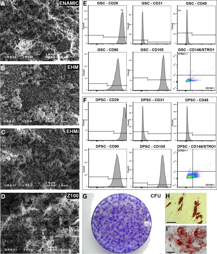
Fig 1. Biomaterials and stem cells characteristics.
SEM microphotographs of the powdered materials. Enamic (Fig 1A), EHM (Fig 1B), EHMi (Fig 1C) and Z100 (Fig 1D). FACS analysis of classical mesenchymal stem cells markers (CD29, 90, 105, 146 and STRO1), of hematopoietic cells (CD45) and of endothalium (CD31) for GSC (Fig 1E) and DPSC (Fig 1F). CFU-F assays of the GSC (Fig 1G). Differentiation staining of the GSC after 14 days: Oil Red O staining (Fig 1H) and Alizarin Red S staining (Fig 1I).Scale bar 100μm.

