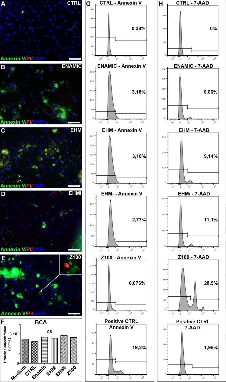
Fig 3. Apoptosis and cell death in contact with the biomaterials.
Detection of apoptosis and cell death. Annexin V and propidium iodide fluorescent stainings of the GSC cultured with no materials (Fig 3A), Enamic (Fig 3B), EHM (Fig 3C), EHMi (Fig 3D) and Z100 (Fig 3E). BCA measurement of the protein concentration in the respective culture conditions (Fig 3F). Flow cytometry analysis of the apoptosis marker Annexin V (Fig 3G) and of dead cells 7-AAD (Fig 3H). Scale bar 100μm.

