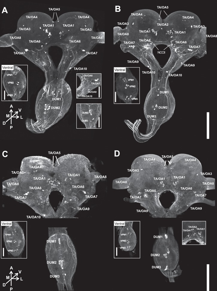
Fig 2. Localization of TA-like-immunoreactive (TA-l-ir) and OA-like-immunoreactive (OA-l-ir) neurons.
TA-l-ir neurons in soldiers (A) and pseudergates (B). OA-l-ir neurons in soldiers (C) and pseudergates (D). Left insets indicate the ventral view of SOG. VPM1, 2, 3 are indicated with black, white, grey filled arrowheads, respectively. Right insets indicate images of other individuals. In right inset of (A), white arrowhead is DUM3 neurons. In right inset of (D), white arrowhead is TA/OA2 neurons. Scale bars indicate 500μm in main picture, and 250μm in insets.

