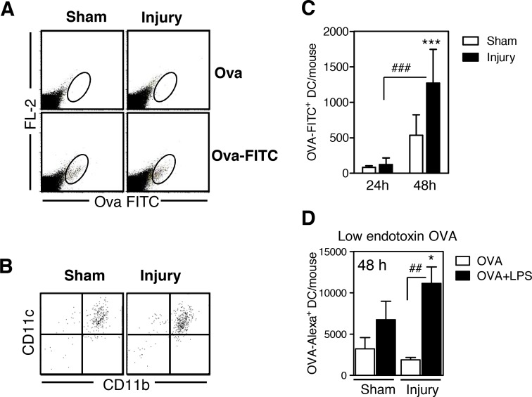
Fig 3. Dendritic cell-like cells in the injured muscle tissue take up ovalbumin after intramuscular application in vivo and migrate into the popliteal lymph node.
Seven days after injury or sham treatment, unlabeled ovalbumin (OVA; Sigma), OVA-fluorescein isothiocyanate (FITC), or OVA-Alexa Fluor 647 was injected into the gastrocnemius muscles. After 24 or 48 h, total popliteal lymph node cells from individual mice were stained for CD11c and CD11b. The FL-2 channel remained free. (A) Representative dot plots of popliteal lymph node cells showing the gating strategy of OVA-FITC+ cells among total lymph node cells 48 h after OVA application. (B) CD11c and CD11b expression of gated OVA-FITC+ cells. (C) Absolute number of OVA-FITC+ dendritic cells (DCs) in the lymph nodes per mouse 24 h (n = 4 per group) and 48 h (n = 9 per group) after injury or sham treatment. (D) OVA-Alexa Fluor 647 alone or in combination with lipopolysaccharide (LPS) was injected, and the absolute number of CD11c+OVA-Alexa+ DCs in the lymph node was determined 48 h later (n = 4 per group). Data are presented as mean±SD. Symbols indicate statistical differences that were detected with analysis of variance (ANOVA). *, p<0.05; ***, p<0.001 sham treatment vs. injury. ##, p<0.01; ###, p<0.001.

