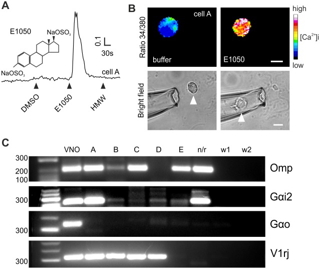
Fig 1. Single-cell RT-PCR following Ca2+ imaging and cell isolation.
(A) Representative intracellular Ca2+ increase (F340/380 ratio, arbitrary units) of a VSN (cell “A”) loaded with fura-2 in response to the sulfated steroid E1050 (chemical structure shown on the side), but not to urine high molecular weight fraction (HMW). (B) Ratiometric (340/380) imaging of the cell shown in A during stimulation with control buffer (DMSO) and E1050. Responsive cells are later picked using a glass capillary micropipette. Arrowhead points to the cell before and after (inside of the micropipette) picking. Scale bar, 10 μm. (C) Ethidium-bromide stained agarose gels of RT-PCR products generated from 5 cells (A to E) showing Ca2+ responses to E1050, a single cell lacking responses (n/r), and two water controls (w1 and w2). cDNA collected from pooled whole VNOs was used as positive control (VNO). PCR amplification of cDNA collected from single cells was performed using gene specific primers for Omp, Gnao1 (Gαo), Gnai2 (Gαi2) and degenerate primers for three members of the V1rj family.

