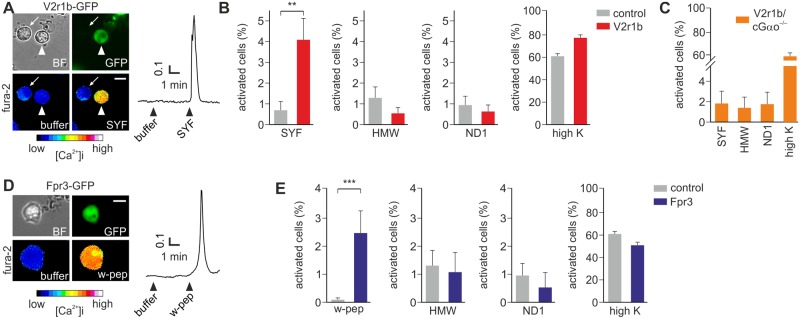
Fig 4. Viral transduction of V2r1b and Fpr3 receptors in VSNs.
(A) Bright field (BF), GFP and pseudocolor fura-2 images of a VSN infected with HSV-V2r1b-IRES-GFP (arrowhead) and activated by the MHC binding peptide SYFPEITHI (SYF). The neighboring non-infected GFP-negative cell (arrow) does not show any calcium increase during peptide stimulation. (B) Summary of cell responses to different stimuli. A 6-fold increase of responsivity to SYF is observed in V2r1b-GFP cells (p < 0.005, Student t test), but not to HMW fraction, the mitochondria-derived peptide ND1, or high K+. (C) Enhanced responsivity to SYF is not observed in Gαo-deficient mice (cGαo-/-) VSNs infected with HSV-V2r1b-IRES-GFP. (D) A single HSV-Fpr3-IRES-GFP infected VSN activated by the synthetic hexapeptide W-peptide (w-pep) is shown. (E) Fpr3-GFP cells show a significantly enhanced number of responses to W-peptide versus GFP control cells (p < 0.001, Student t test), but not to HMW, ND1 or high K+. N = 469 V2r1b-GFP, 163 Fpr3-GFP and 1224 control-GFP cells, in 12, 8 and 27 experiments, respectively. Scale bars, 10 μm.

