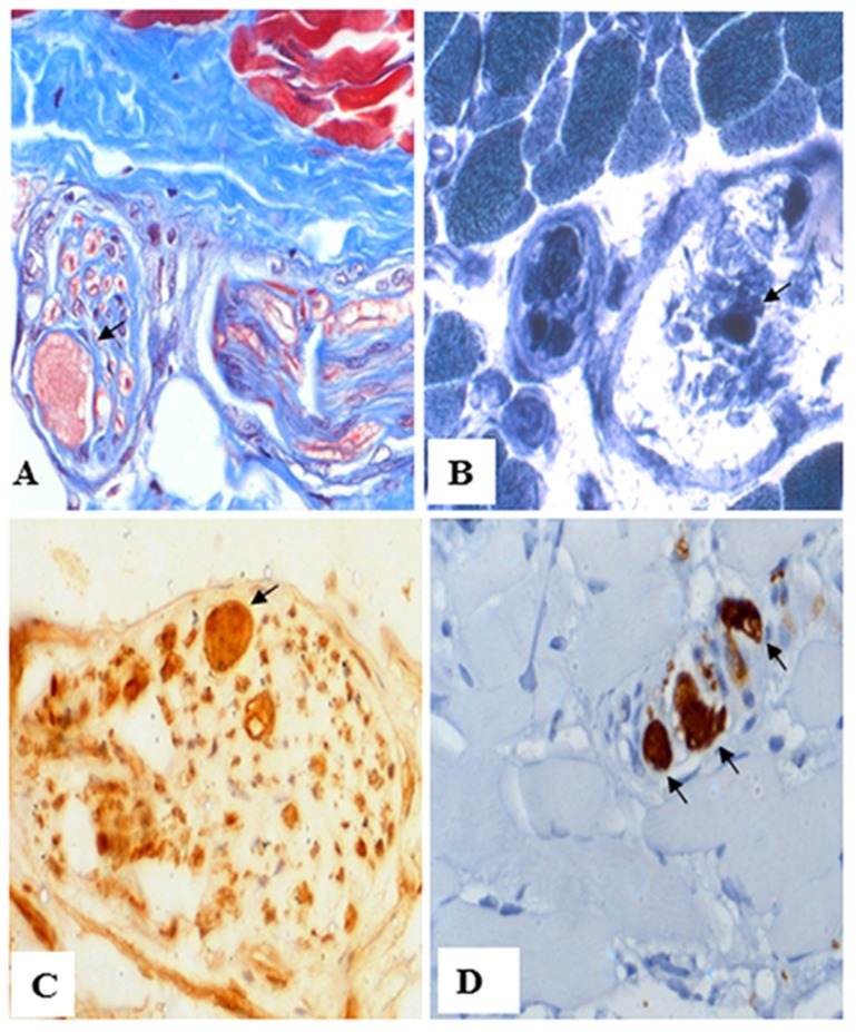
Fig 2. Muscle nerve pathology of affected individual II-1 from family 7.
(A) Muscle biopsy reveals marked axonal distension of intramuscular nerve twig (arrow), using Masson's trichrome staining. (B) The intraaxonal contents in the distended axon contain NADH-TR (arrow). (C) Phosphorylated neurofilament staining (arrow). (D) Ubiquitin staining (arrow). Magnifications: (A) 200X; (B) 200X; (C) 400X; and, (D) 200X.

