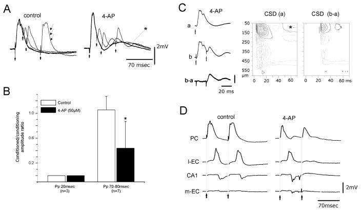
Fig. 2.
(A) Paired-pulse (Pp) stimulation with different ISIs induces suppression of disynaptic potentials in the conditioned PC response. The disynaptic component (arrows) recovers at an ISI of 70 ms under control conditions. During perfusion with 4AP the paired-pulse depression of disynaptic potential did not recover at an ISI of 70 ms. The asterisk marks the delayed slow potential induced by 4AP in the PC. (B) Ratio between the amplitudes of the conditioned (second) and the conditioning (first) disynaptic components at ISIs of 20 and 70–80 ms in control solution (gray columns) and during 4AP application (black columns). (C) PC response to single stimulation (a) and to paired-pulse stimulation at 20 ms ISI (b), and digitally subtracted trace (b–a). The CSD contour plot of the b–a laminar profile shows that only the monosynaptic components located in the most superficial cortex remain after the conditioned stimulus. Isocurrent lines at 0.14 mV / mm2. (D) Field potentials simultaneously recorded in the PC, l-EC, CA1 and m-EC after double-pulse stimulation of the LOT in control condition and during 4AP perfusion. 4AP blocks the propagation of activity evoked by the second stimulus of the pair.

