


Fig. 2.
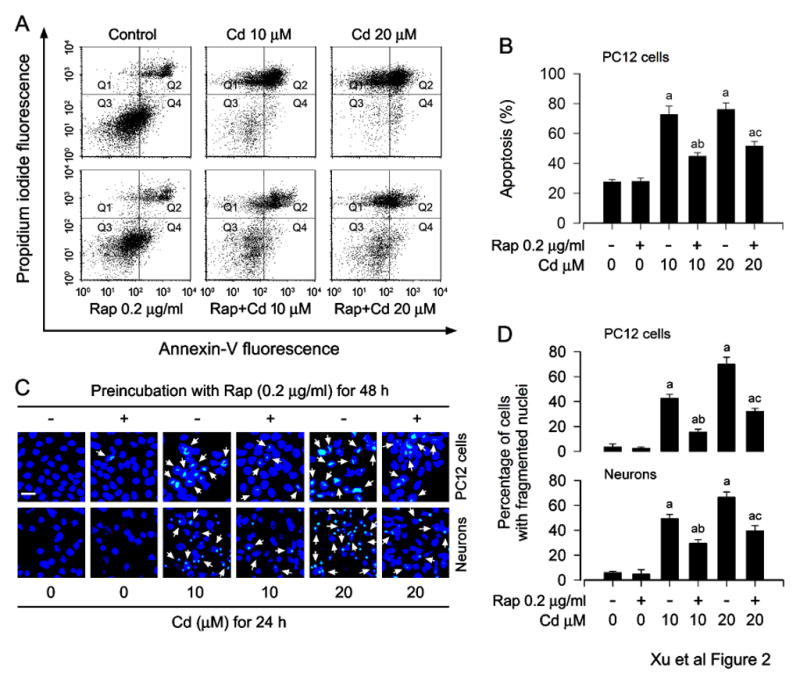
Rapamycin prevents Cd-induced apoptosis in neuronal cells. PC12 cells and/or primary neurons were pretreated with rapamycin (Rap, 0.2 μg/ml) for 48 h, and then exposed to Cd (10 and 20 μM) for 24 h. A) The percentages of live (Q3), early apoptotic (Q4), late apoptotic (Q2) and necrotic cells (Q1) were determined by FACS using annexin-V-FITC/PI staining. The results from a representative experiment are shown. B) Quantitative analysis of apoptotic PC12 cells by FACS assay. C) Apoptosis in PC12 cells and primary neurons was evaluated by nuclear fragmentation and condensation (arrows) using DAPI staining. Scale bar: 20 μm. D) The percentages of cells with fragmented nuclei were quantified, showing that rapamycin markedly attenuated Cd-induced apoptosis in the cells. Results are presented as mean ± SEM (n = 5). a p < 0.05, difference with control group; b p < 0.05, difference with 10 μM Cd group; c p < 0.05, difference with 20 μM Cd group.