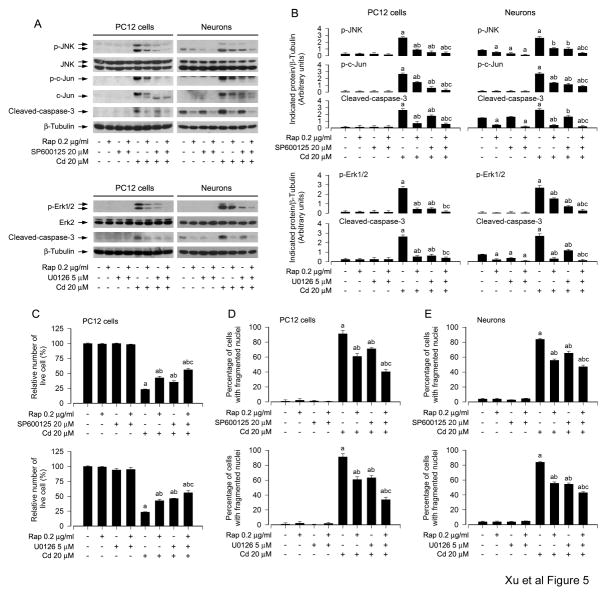
Fig. 5.
Rapamycin inhibits Cd-induced neuronal apoptosis by blocking JNK and Erk1/2 pathways. PC12 cells and primary neurons were pretreated with/without rapamycin (0.2 μg/ml) for 48 h, and then SP600125 (20 μM) or U0126 (5 μM) for 1 h, followed by exposure to Cd (20 μM) for 4 h (for Western blotting) or 24 h (for live cell analysis, DAPI staining). A) Cell lysates were subjected to Western blot analysis using indicated antibodies, showing that inhibitors of JNK (SP600125) and Erk1/2 (U0126) strengthened the inhibitory activity of rapamycin. The blots were probed for β-tubulin as a loading control. B) Similar results were observed in at least three independent experiments, and blots for p-JNK, p-c-Jun, p-Erk1/2, and cleaved-caspase-3 were semi-quantified. C) Live cells were detected by counting viable cells using trypan blue exclusion and D and E) the percentages of apoptotic cells with fragmented nuclei were quantified by DAPI staining, showing that pharmacological inhibition of JNK and Erk1/2 enhanced rapamycin prevention of Cd-induced cell viability reduction and apoptosis, respectively. Results are presented as mean ± SEM (n = 3–5). a p < 0.05, difference with control group; b p < 0.05, difference with 20 μM Cd group; c p < 0.05, difference with Cd/SP600125 group, Cd/U0126 group or Cd/Rapamycin group.

