Figure 3.
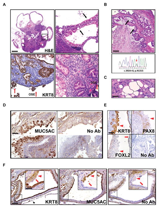
Mucinous epithelial ovarian tumors develop in PKP53H/+ mice. A, At 12 weeks of age, 80% of PKP53H/+ mice develop mucinous structures and some of them become cystic (top panel). Regions in frame are shown at higher magnification at the right. Red arrows: OSE cells. Black arrows: secretory mucinous structures. Scale bar: 900μm, 180 μm and 90 μm. B, Representative image of H&E stained human mucinous EOC tumor (upper panel) and the corresponding sequencing results of the TP53 gene in laser capture microdissected tumor cells from the same patient. Black arrows: secretory mucinous structures. Scale bar: 200 μm. C, The RMUG-L human cell line, derived from a mucinous ovarian carcinoma, carries heterozygous WT and mutantTP53 alleles and exhibits secretory mucinous structures when innoculated into the intraperitoneal cavity of SCID mice. Scale bar: 25 μm. D, Mucinous structures in PKP53H/+ mice express MUC5AC, a marker for human mucinous ovarian carcinoma. Adjacent sections without first antibody were used as negative controls. Scale bar: 100 μm. E, Mucinous structures in PKP53H/+ mice express KRT8 but do not express FOXL2 (nuclear staining) or PAX8 (nuclear staining). Red arrowheads: mucinous structures; red arrows: epithelial structures. Scale bar: 150 μm. F, Some cystic structures in the ovary of PKP53H/+ mice contain contiguous regions that are positive for both MUC5AC and KRT8 (red arrow heads) or for KRT8 alone (red arrows) (inserts are higher magnification of regions in frames). Black arrowhead: ovarian bursa. Scale bar: 200 μm and 100 μm.

