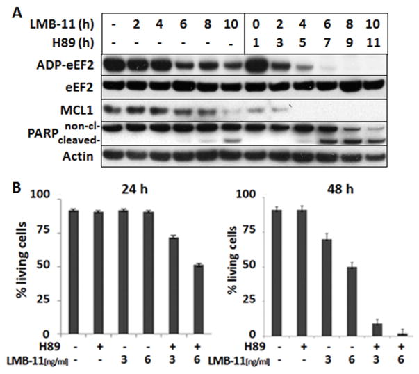
Figure 3. H89 increased ADP-ribosylation of eEF2 and rate of apoptosis.
A. KOPN-8 cells were treated with 10 μM H89 for 1 hour, then, 200 ng/ml of LMB-11 was added for the indicated times before cell lysis, followed by Western Blot analysis for Mcl-1, cleaved PARP, and actin. Not yet modified eEF2 in the cell lysates was labeled in a cell free reaction with biotinylated ADP by LMB-11 as described in methods. B. KOPN-8 cells were treated with 10 μM H89 or vehicle for 1 hour, then left untreated or treated with the indicated concentrations of LMB-11 for 24 (left panel) or 48 hours (right panel), respectively. Cells were then stained with Annexin V-PE and 7-AAD. Living cells were defined as double negative and blotted as percentage of all cells. Standard errors of mean were generated from three independent experiments.

