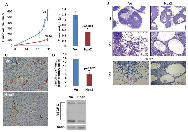
Figure 1.
Hpa2 over expression attenuates tumor growth. Control (Vo) and Hpa2 over expressing FaDu cells (5×10⁶) were implanted subcutaneously in SCID mice and tumor volume was inspected (A, left). At termination, tumor xenografts were collected, weighed (A, right) and formalin-fixed. Paraffin-embedded 5 micron sections were subjected to histological examination. Shown are representative images of hematoxylin & eosin (H&E) staining at low (B, upper panel) and high (B, middle panel) magnifications. H&E staining of tumor xenografts produced by control (Vo) and Hpa2 over expressing Cal27 oral carcinoma cells is shown in B, lower panels. Note massive necrosis in control tumors vs. cysts structures in tumors over expressing Hpa2. Original magnifications: Upper panels ×4, middle and lower panels ×10. C–D. Lymph angiogenesis and VEGF-C expression. Five micron sections of tumor xenografts produced by control (Vo) and Hpa2 over expressing FaDu cells were stained with anti-LYVE-1 antibody, a marker for lymphatic endothelial cells (C); quantification of lymph vessel density is shown graphically in D (upper panel). Original magnification: ×40. Extracts of control and Hpa2 over expressing cells were subjected to immunoblotting applying anti-VEGF-C and anti-actin antibodies (D, lower panels).

