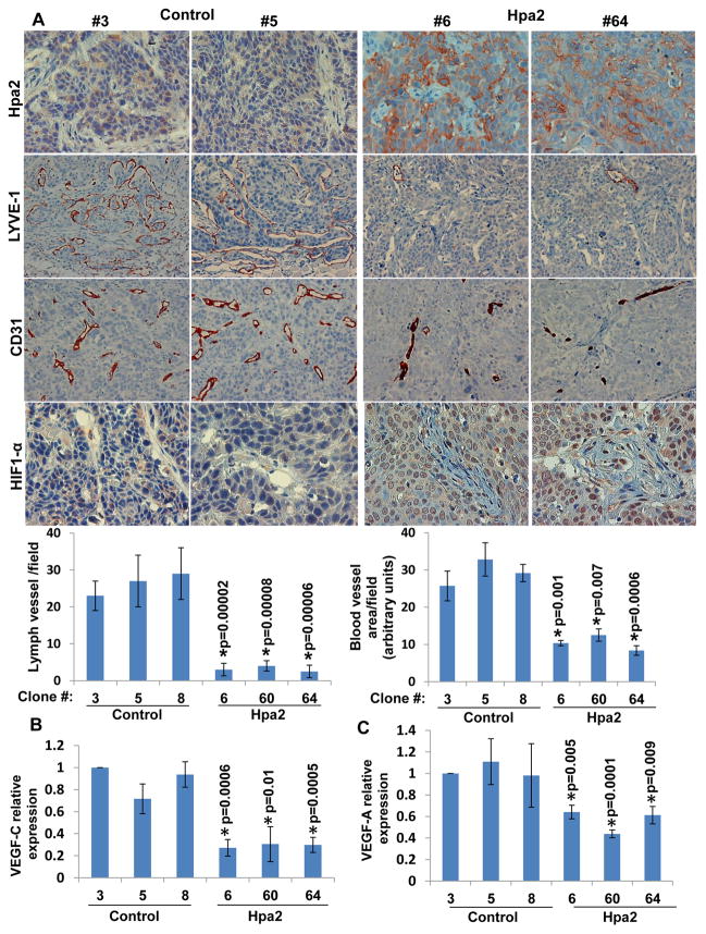
Figure 3.
Hpa2 over expression is associated with decreased tumor vascularity. A. Immunostaining. Sections of tumor xenografts produced by control (#3, #5) and Hpa2 over expressing (#6, #64) cell clones were subjected to immunostaining applying anti-Hpa2 (upper panels), anti-LYVE-1 (second panels), anti-CD31 (third panels), and anti-HIF-1α (fourth panels) antibodies. Note, reduced tumor vascularity and increased tumor hypoxia following Hpa2 over expression. Original magnifications: upper, third and fourth panels ×40, second panels ×20. Quantification of lymph (LYVE-1-positive) and blood (CD31-positive) vessels is shown graphically in the lower panels. B–C. Real time PCR. Total RNA was extracted from the indicated cell clones and corresponding cDNAs were subjected to quantitative real-time PCR analyses. Expression of VEGF-C (B) and VEGF-A (C) are presented graphically in relation to the levels in control clone #3, set arbitrarily to a value of 1.

