Figure 5.
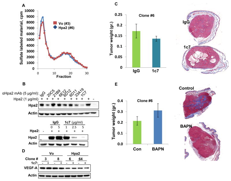
Hpa2 mode of action. A. Heparanase activity. Control (#3) and Hpa2 over expressing (#6) cell clones were plated on dishes coated with sulfate-labelled ECM and grown for three days. Conditioned medium was then collected and evaluated for heparanase activity as described in ‘Materials and Methods’. B. Characterization of monoclonal antibody (mAb) 1c7. Hpa2 (1 μg/ml) was added to 293 cells together with the indicated anti-Hpa2 mAb or control mouse IgG (5 μg/ml; 30 min, 37ºC) under serum-free conditions. Medium was then aspirated, cells were washed twice with PBS and lysate samples were subjected to immunoblotting applying anti-Hpa2 (upper panel) and anti-actin (second panel) antibodies. Decreased cellular binding of Hpa2 by mAb 1c7 is shown to be dose-dependent (third panel). C. Tumor xenografts. Hpa2 over expressing clone #6 cells were implanted (5×10⁶) subcutaneously in SCID mice (n=7) and mice were administrated with mAb 1c7 (250 μg/mouse) three times a week. At termination, tumors were collected, weighed (left panels) and processed for histological examination. Shown are representative images of whole sections stained with Masson’s/Trichrome reagents and scanned by 3DHISTECH Pannoramic MIDI System attached to HITACHI HV-F22 color camera (3dhistech kft, Budapest, Hungary). Note that mAb 1c7 does not affect tumor growth but appears to enhance cyst formation. D. VEGF-A expression. The indicated cell clones were incubated without (-) or with (+) mAb 1c7 (5 μg/ml, 18 h, 37ºC) and lysate samples were subjected to immunoblotting applying anti-VEGF-A (upper panel) and anti-actin (lower panel) antibodies. Note decreased VEGF-A levels in cells over expressing Hpa2, which is not affected by mAb 1c7. E. LOX inhibitor. SCID mice (n=7) were inoculated subcutaneously with Hpa2 over expressing clone #6 cells (5×10⁶) and mice were administrated with LOX inhibitor BAPN (0.75% in drinking water). At termination, tumors were collected, weighed (left panels) and processed for histological examination. Shown are representative images of whole sections stained with Masson’s/Trichrome reagents and scanned by 3DHISTECH Pannoramic MIDI System (right panels). Note that LOX inhibitor does not affect tumor growth but reduces tumor fibrosis (i.e., collagen content).

